
Photographs from the collections of
Museo di Anatomia Umana “Filippo Civinini”
Università di Pisa
April 2019
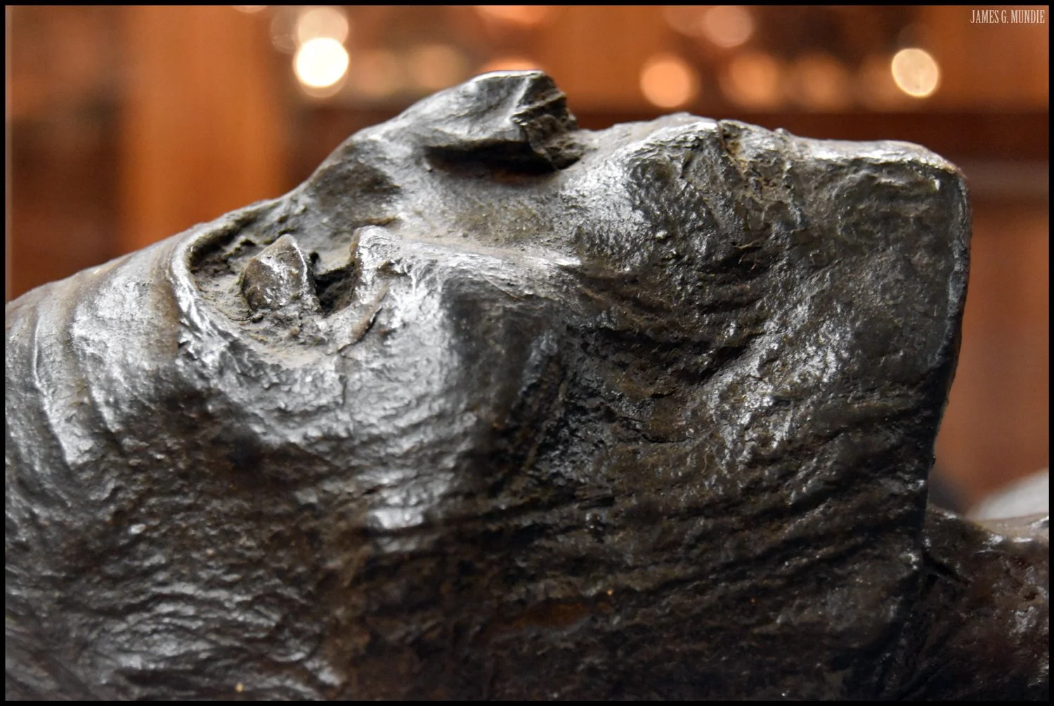
Egyptian mummy
Officially established in 1343, the Università di Pisa is one of the oldest universities in Europe, with evidence suggesting its origins date back to the 11th century. After a period of decline in the 14th and 15th centuries—including a nine-year relocation to Florence—the university experienced a revival under the patronage of Cosimo I de’ Medici (1519–1574). During his leadership, a medical school was founded and an anatomical theater constructed. It was in this setting that Andreas Vesalius (1514–1564), along with many other prominent anatomists, conducted dissection lectures.
This rich academic legacy laid the foundation for the Museo di Anatomia Umana in Pisa. The museum project began in 1832 when professor of anatomy Tommaso Biancini started assembling what would become the Anatomical Cabinet. The task of organizing and cataloging the collection was later taken over by Filippo Civinini (1805–1844), then chair of anatomy, for whom the museum is now named. The museum opened to the public in 1834.
By the mid-19th century, the collection had grown to over 1,000 items and expanded to include ancient Egyptian and pre-Columbian funerary objects, including mummies. Today, the museum houses more than 3,000 anatomical preparations, casts, models, illustrations, and related artifacts.
Special thanks to Professor Gianfranco Natale and the staff of Museo di Anatomia Umana for providing access to the collections and their generous hospitality.
A close-up, black-and-white image of a preserved human head with a sticker on the side, reflecting light and creating a hazy effect.

A human skull with the top surface marked with handwritten Latin words and numbers in circles, likely representing parts of the skull and brain regions.

A black and white eerie image showing a skull with an eyeball in its eye socket, covered in small white particles, with a classified card labeled '7' and a partially visible sign in the background reading 'ANATOMICO' and 'PISA'.

A group of human skeletons displayed together, with skulls and bones visible, in black and white.

Close-up of a human skull with handwritten labels and markings on the bone surface.

Close-up of the face of an Egyptian mummy, lying down, facing upward with closed eyes.

Close-up black-and-white photograph of a mummifieded face, showing texture and cracks in the tissue.

An anatomical illustration of a human male with veins and arteries visible throughout the body, with the right arm raised and bent.

An anatomical chart of the human body showing muscles, bones, and the circulatory system, with muscles colored red, veins shown in blue, and arteries in red.

Example of blurred text tattooed on preserved human skin, stretched onto a wire support from for display in a musuem.

Close-up of an expaneded human skull with screws and wires attached, and separated bones and anatomical features visible.

Close-up of a glass container containing a partially dissected face preserved in a yellowish-greenish liquid, with a label indicating it was from Pisa.

A presevered human fetus inside a transparent container for display in a museum.

Close-up of conjoined twins submerged in a jar of preservative liquid on display in a museum.

A group of human skeletons and skulls arranged closely together in a display, rendered in black and white.

A large glass container with a human face, showing one eye partially open, with a label in Italian, 'STUDIO ANATOMICO' and 'PISA', and a smaller label with the number 24.

Close-up of a large glass container with a human face, showing one eye partially open.

A human skull with hand-drawn diagrams and text on the top, showing circles and Italian names of regions of the brain or head.

A preserved fetus submerged in a glass container filled with preservative liquid.

Close-up of two preserved human fetuses in an anatomical display case.
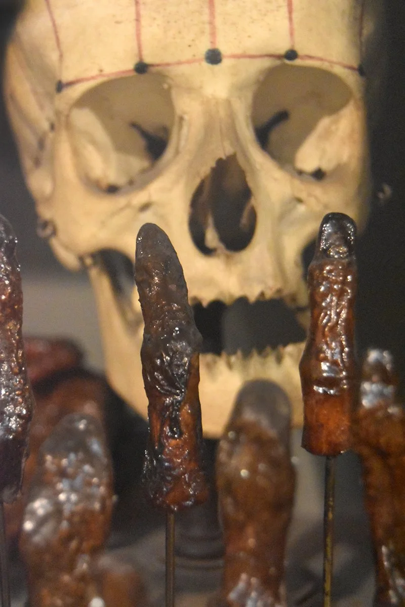
A human skull with several preserved human fingers front of it.

A preserved human skull with decayed flesh and exposed bones.

A close-up of a wax anatomical sculpture of a human face with yellowish skin tone, shaowing a partially dissected torso.

An anatomical model of a human body showing muscles, nerves, and organs.

A glass jar with a handwritten note inside, dated 15th August 1869, describing the location of a wooden cross and some instructions in cursive handwriting.
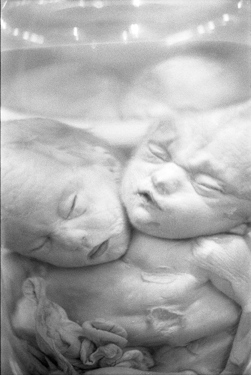
A black and white photograph of preserved conjoined twins.

A glass jar containing a preserved reddish-orange specimen. Attached to the jar is an old handwritten note in Spanish, likely describing the specimen and its collection details.

Four human skulls displayed side by side in black and white.

Ancient mummified human skull with decayed teeth and remnants of skin and tissue.

Close-up of a weathered, mummified face with closed eyes, brown skin, and brown hair, surrounded by animal fur.

A preserved head inside a glass display case, labeled with a small sign showing the number 22, in an exhibit about Pisa.

Close-up of a human skull with handwritten notes and drawings etched on its surface.

A human skull replica with a detailed brain model attached, displayed inside a glass case.

A detailed anatomical model of the human neck and head, showing muscles, blood vessels, and nerves, with the skin removed to expose internal structures.

Close-up of a preserved human cadaver for anaotmical study, with hollow eye sockets and exposed teeth.

Two glass beakers containing preserved biological specimens submerged in liquid, with visible writing on the beakers, in an indoor setting.

Conjoined twins preserved in a plastic container.

A preserved human fetus inside a glass jar and a mummified human skull and torso on display in a museum.
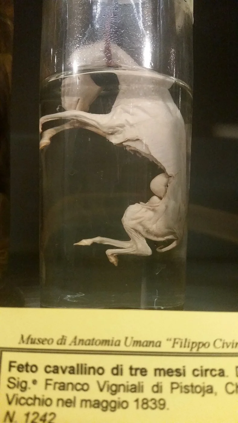
A preserved fetal horse specimen in a glass container, from Museo di Anatomia Umana. The specimen is approximately three months old, from the Pistoja region, collected in May 1839.

Several human skeletons with skulls on display in a museum.

A close-up of a preserved human cadaver with large eyes, prominent nose, and uneven teeth.

An anatomical illustration of the human body showing muscles, arteries, and nerves, with a focus on the head, upper body, and arms.

Illustration of human anatomy focusing on the head and neck, showing muscles, blood vessels, and structural details.

A preseved human skull and partially skeletal hand, displayed in a museum setting.

A preserved dissected cadaver with an open mouth showing teeth and hand raised. The background appears to be a dimly lit museum or gallery with display cases.

A preserved human skeleton with a skull and ribcage, displayed in a museum case.

Mummified human skull with a tag hanging from a string.

A mummified human skull with missing teeth and darkened areas, mounted against a gray background.

A mummified human body with a gaunt face and missing teeth, sitting with one legs drawn to the torso.

Close-up photograph of a Peruvian mummy.

Preserved fetal speciman, with eyes half-closed and mouth open in an expression that appears to be yawning or shouting, set against a blurred background.

Ancient Egyptian funerary painting depicting figures and objects in profile, some holding staffs or ritual items.

Close-up of a ceramic jar in the form of a face with large round eyes, open mouth, and a grid pattern on the face. The surface appears weathered and aged.

Close-up of a carved sculpture of a human face with wide eyes and small hands on the chest.
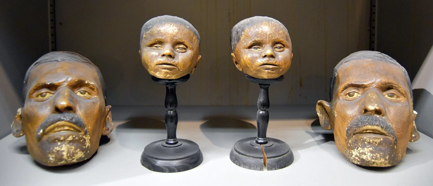
Four antique human skull and head sculptures, two with gemstone earrings, some with gold leaf. The skulls and heads are placed on a white surface in a display case.

A preserved dissected fetus with the head and upper body visible, encased in a glass display case.

Front view of a historic building labeled 'SCUOLA MEDICO-CHIRURGICA', featuring arched windows, a central entrance, and flags of the European Union, Italy, and a third country or organization, with a person sitting near the entrance and the building surrounded by a metal fence.